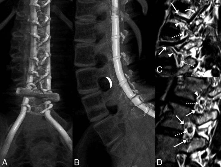
Fig 2.
A and B, CT volumetric rendering of a patient with SMA2 demonstrates extensive posterior spinal fusion hardware and complete osseous interlaminar fusion without any access for a classic interlaminar LP. Note the widely patent neural foramina. The white crescent represents the target for TFLP. C and D, Sagittal 3D volumetric T2-weighted images of a healthy person obtained with 3T MR imaging. C, Image obtained slightly lateral to the neural foramen. D, Image obtained at the foramen. Flow voids of the lumbar arteries (arrowheads) and larger caliber lumbar veins (arrows) are seen in the anterior superior aspect of the foramen. A branching ascending lumbar vein is seen coursing toward a higher level neural foramen (curved arrow). More venous structures are seen in the inferior aspect of the foramen (arrows in D). Exiting nerve roots are shown within the center of the foramen (dashed arrows).

