Fig 4.
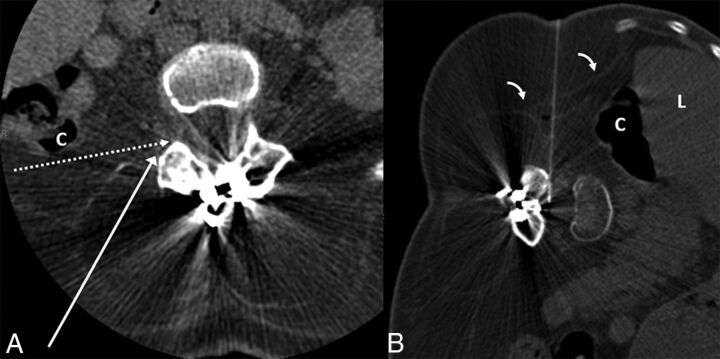
A 36-year-old woman with SMA2 (case 6). A, Preprocedural CT obtained in supine position at the level of L3–4 demonstrates extensive muscle atrophy. The long white arrow shows the normal needle trajectory for a transforaminal epidural steroid injection. The dashed white arrow indicates the needle trajectory for TFLP. Note that while supine, the posterior margin of the ascending colon (C) is along the proposed needle trajectory. B, CT fluoroscopy image during TFLP obtained with the patient in the left lateral decubitus position. The needle is advanced into the thecal sac with an angle slightly >90°, just anterior to the facet. Note the anterior displacement of the liver (L) and ascending colon (C) in the decubitus position, providing a safer approach. Of note, the thin transversalis fascia (bent arrows) is well-visualized.

