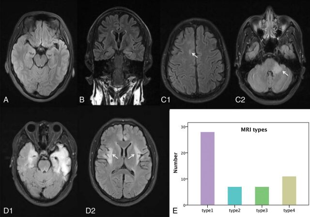
Fig 1.
Four types of brain MR imaging appearances in patients with anti-NMDA receptor encephalitis, and the histogram of the 4 types of brain MR imaging appearance. Axial (A, C, and D) and coronal FLAIR images (B) come from 4 patients (C1 and C2 from same patient, D1 and D2 from same patient). A, Type 1, a 23-year-old male patient with anti-NMDA receptor encephalitis, with normal brain MR imaging findings. B, Type 2, a 29-year-old female patient. Lesions are in the left hippocampus with bilateral mild volume loss in the hippocampus. C, Type 3, a 28-year-old male patient. Lesions are in the right frontal lobe (white arrow) and middle cerebellar peduncle (white arrow) and brain stem. D, Type 4, a 25-year-old male patient, with lesions located in the bilateral frontal lobe, temporal lobe, insula (white arrows), hippocampus, and cingulate gyrus, with volume loss in the left hippocampus. E, Histogram of the 4 types of brain MR imaging appearances.

