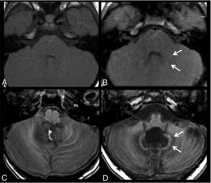
Fig 2.
T1-weighted images acquired before (A and C) and after 20 gadobenate injections (B and D) in a patient with follow-up for optic glioma without radiation or chemotherapy (A and B) and a patient with medulloblastoma after an operation and radiochemotherapy (C and D). Subtle signal changes of the dentate (arrows) can be seen in the patient without any therapy, while the patient with RCTX shows distinct T1 signal changes of the dentate and perifocal edema.

