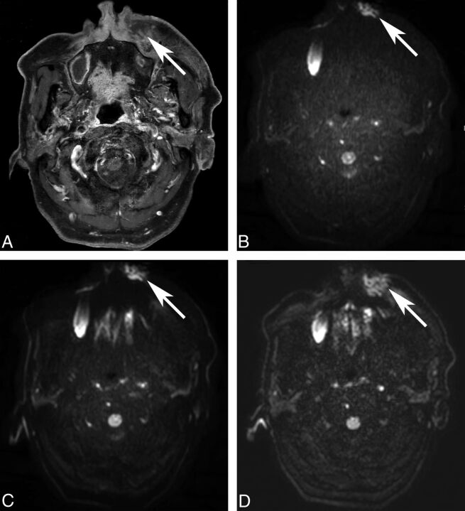
Fig 2.
Image distortion of DWI. An 85-year-old man underwent MR imaging for the evaluation of a left premaxillary abscess (A, post-contrast-enhanced T1WI, arrow). Image distortion is increased on ssEPI-STIR (B, arrow) and ssEPI-SPIR (C, arrow) and decreased on msEPI-SPIR (D, arrow). The left premaxillary abscess is not distorted by the susceptibility artifacts only on msEPI-SPIR.

