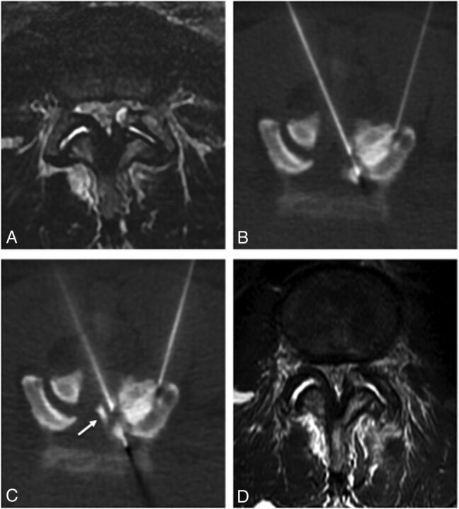
Fig 1.
Successful CT-guided cyst puncture, aspiration, and fenestration. A, Axial T2-weighted MR image demonstrates bilateral L4–5 facet synovitis and a thin-rimmed T2-hyperintense cyst arising from the left L4–5 facet joint. B, Intraprocedural CT image shows contrast opacification of the cyst via injection into a 22-ga spinal needle placed within the left L4–5 facet joint (step 1). A second 22-ga spinal needle has been advanced coaxially into the cyst via a translaminar approach (step 2). C, CT image obtained after cyst aspiration and repeat fenestration demonstrates successful cyst perforation with leakage of contrast into the epidural space (arrow, step 3). D, Follow-up MR imaging shows resolution of the treated cyst.

