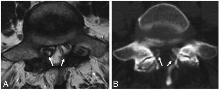
Fig 2.
Calcified synovial cysts. A, Axial T2-weighted MR image shows severe bilateral L5–S1 facet arthrosis with thick-rimmed facet cysts (arrows), which result in severe lateral recess narrowing on the right and indentation of the thecal sac on the left. B, Axial CT image shows peripheral calcification of both cysts (arrows). Direct cyst puncture and fenestration were not performed in this patient, who instead underwent CT-guided facet injections and nerve blocks and ultimately required an operation.

