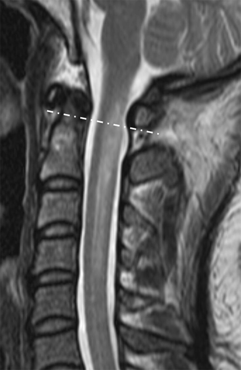
Fig 1.

Sagittal T2-weighted spinal MR imaging of a 36-year-old woman with LETM. The lesion extends beyond the imaginary line (dashed line) connecting the inferior cortex of the C1 anterior and posterior arches. Cervicomedullary junction involvement is present.
