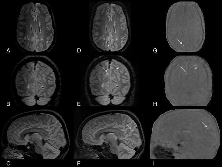
FIGURE.
Detection of leptomeningeal contrast-enhancement foci using subtraction images. A–C, Coregistered 3D-FLAIR precontrast images in all 3 orthogonal planes. D–F, The corresponding coregistered 3D-FLAIR postcontrast images in all 3 orthogonal planes. G–I, The corresponding pre-/postcontrast 3D-FLAIR subtraction images in all 3 orthogonal planes. A patient with relapsing-remitting multiple sclerosis has a true LM CE in the right parietal region that was easily spotted with the aid of pre-/postcontrast 3D-FLAIR subtraction images, which otherwise would have been undetected.

