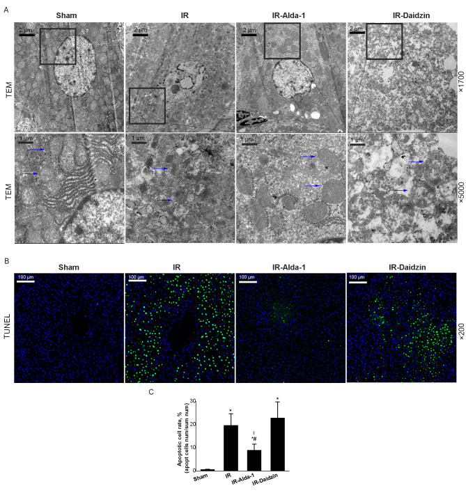
Figure 5.
Alda-1 pretreatment reduces damage to liver mitochondria and hepatocyte apoptosis in HIRI model rats. (A) Representative transmission electron micrographs showing mitochondria in the liver tissue after 24 h of reperfusion. Blue arrows indicate mitochondria. Magnification, ×1,700 and ×5,000. Scale bars, 1 or 2 µm. (B) Representative TUNEL-stained images showing HIRI rat livers after 24 h of reperfusion. Magnification, ×200; scale bar, 100 µm. (C) TUNEL-stained apoptotic cell rate of each group. Data are presented as the mean ± SD. n=6. *P<0.05 vs. the sham group; #P<0.05 vs. the IR group; $P<0.05 vs. the IR-Daidzin group. RP, reperfusion; HIRI, hepatic ischemia/reperfusion injury; IR, ischemia/reperfusion; TEM, transmission electron microscope.

