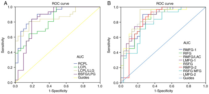
Figure 5.
ROC curve analysis of the mean ALFF values of the altered brain regions in VH. (A) The areas under the ROC curve were: RCPL 0.893, (P<0.001; 95% CI, 0.814–0.972), LCPL 0.823 (P<0.001; 95% CI, 0.723–0.923), LCPL/LLG 0.856 (P<0.001; 95% CI, 0.761–0.951) and BSFG/LPG 0.839 (P<0.001; 95% CI, 0.732–0.945). (B) The areas under the ROC curve were: RMFG-1 0.847 (P<0.001; 95% CI, 0.748–0.946), RIFG 0.837 (P<0.001; 95% CI, 0.739–0.934), RMFG/LAC 0.890 (P<0.001; 95% CI, 0.807–0.972), LMFG-1 0.868 (P<0.001; 95% CI, 0.782–0.954), RSFG 0.864 (P<0.001; 95% CI, 0.773–0.954), RMFG-2 0.832 (P<0.001; 95% CI, 0.724–0.941), RSFG/MFG 0.781 (P<0.001; 95% CI, 0.666–0.897) and LMFG-2 0.850 (P<0.001; 95% CI, 0.754–0.947). ROC, receiver operating characteristic; ALFF, amplitude of low-frequency fluctuation; RCPL, right cerebellum posterior lobe; CI, confidence interval; LCPL, left cerebellum posterior lobe; LLG, left lingual gyrus; BSFG/LPG, bilateral superior frontal gyrus/left postcentral gyrus; RMFG, right middle frontal gyrus; RIFG, right inferior frontal gyrus; LAC, left anterior cingulate; LMFG, left middle frontal gyrus; RSFG, right superior frontal gyrus; MFG, middle frontal gyrus; AUC, area under the curve.

