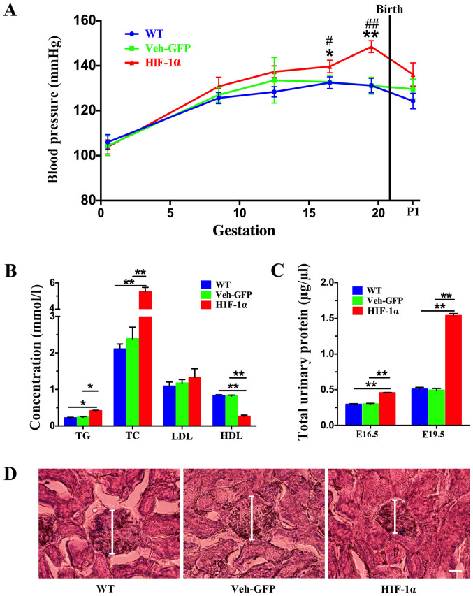
Figure 1.
Construction of a PE mouse model by Hif-1α virus. PE mice exhibited elevated blood pressure, increased urinary protein levels and impaired renal function. (A) Blood pressure of the WT, Veh-GFP and Hif-1α mice was measured throughout gestation at the indicated time points (n=8 per group). Data are presented as the mean ± SEM from several mice used in each experiment. *P<0.05 and **P<0.01 vs. Veh-GFP group; #P<0.05 and ##P<0.01 vs. WT group.(B) TG, TC, LDL and HDL levels in the serum of WT, Veh-GFP and Hif-1α mice at E19.5 were measured (n=4 per group). Data are presented as the mean ± SEM from several mice used in each experiment. *P<0.05, **P<0.01. (C) Total urinary protein level in WT, Veh-GFP and Hif-1α mice at E16.5 and E19.5 was determined using bicinchoninic acid protein assays (n=8). **P<0.01. (D) Renal histology of WT, Veh-GFP and Hif-1α mice at E19.5 demonstrated by hematoxylin and eosin staining. Scale bar, 50 µm. PE, preeclampsia; Hif-1α, hypoxia-inducible factor 1α; WT, wild-type; Veh-GFP, vehicle-green fluorescent protein; E, embryonic day; TG, triglyceride; TC, total cholesterol; LDL, low-density lipoprotein; HDL, high-density lipoprotein.

