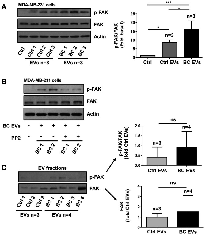
Figure 6.
EVs isolated from patients with breast cancer induce FAK activation via a Src-dependent pathway. (A) Lysates from MDA-MB-231 cells treated for 20 min with three Ctrl EVs and three BC EVs were analyzed by western blotting with anti-p-FAK Ab. Membranes were further analyzed by western blotting with anti-FAK Ab and anti-actin Ab as loading controls. (B) MDA-MB-231 cells were untreated and treated for 1 h with 10 µM PP2 and stimulated for 20 min with two BC EVs and lysed. Cell lysates were analyzed by western blotting with anti-p-FAK Ab. Membranes were analyzed further with anti-FAK Ab and anti-actin Ab as loading controls. (C) Three Ctrl EVs and four BC EVs were analyzed by western blotting with anti-p-FAK Ab and anti-FAK Ab. Data are presented as the mean ± SD, and indicate the fold of p-FAK or FAK above Ctrl and Ctrl EVs. *P<0.05, ***P<0.001 vs. Ctrl and Ctrl EVs. ns, not significant; Ctrl, control; Ab, antibody; EVs, extracellular vesicles; Ctrl EVs, EV fractions obtained from healthy women; BC EVs, EV fractions obtained from women with breast cancer; p-, phosphorylated; FAK, focal adhesion kinase.

