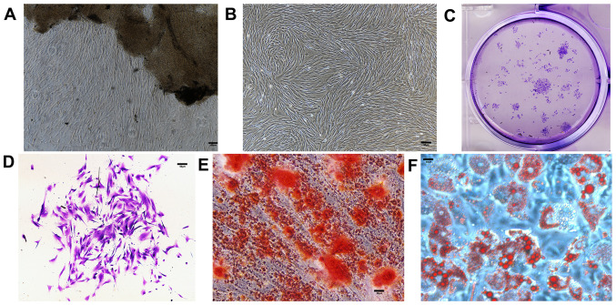
Figure 2.
In vitro characteristics of cultured PDLSCs. Scale bars=200 µm. (A) PDLSCs grew around periodontal ligament tissues five days after incubation. (B) PDLSCs reached confluence seven days after incubation. (C) Entire plate view of colony-forming PDLSCs, which were plated at a low density and cultured for 7 days before staining with crystal violet. (D) Cell clusters derived from PDLSCs with typical fibroblast-like morphology seven days after incubation. (E) Alizarin Red-S staining 14 days after incubation. (F) Oil Red O staining four weeks after incubation. PDLSC, periodontal ligament stem cell.

