


Figure 2. Comparison of the three models of BH3-only mediated Bax/Bak activation.
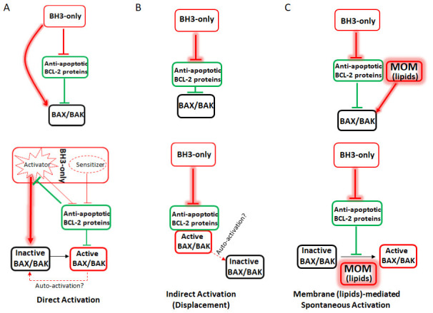
A) The direct activation model classifies BH3-only proteins as either activators (i.e. Bid, Bim, and Puma) or sensitizers (Bad). When not sequestered by anti-apoptotic Bcl-2 proteins, the activators directly bind to and activate Bax/Bak. Sensitizers cannot directly activate Bax/Bak but can bind to anti-apoptotic Bcl-2 proteins. To initiate apoptosis, the sensitizers bind to the anti-apoptotic Bcl-2 proteins, which release the activators from sequestration and allow them to directly activate Bax/Bak. B) The indirect activation model hypothesizes that Bax/Bak are constitutively active but are sequestered by the anti-apoptotic Bcl-2 proteins. During apoptosis, the BH3-only proteins bind to the anti-apoptotic Bcl-2 proteins and displace active Bax/Bak, which then damage the mitochondria. It is also hypothesized that active Bax can recruit inactive cytosolic Bax to the mitochondria through auto-activation. C) The membrane (lipids)-mediated spontaneous activation model hypothesizes that during apoptosis, the BH3-only proteins bind to and neutralize the anti-apoptotic Bcl-2 proteins. This allows the mitochondrial outer membrane (MOM) lipids to serve as the direct activator of Bax/Bak, as Bax freely migrates to the MOM through free diffusion. Helix 9 mediates membrane association for both Bax/Bak, and such association results in spontaneous activation of Bax/Bak in the lipid bilayer of the MOM. Similar to the indirect activation model, the BH3-only proteins’ only function is to inactivate the anti-apoptotic Bcl-2 proteins. However, in the membrane (lipids)-mediated spontaneous (MMS) model, both Bax and Bak are directly activated by the MOM. Figure 2c is adapted from Figure 5b of our previous paper in Cell Research 59.