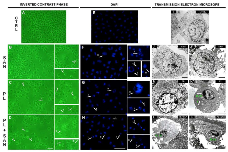
Figure 6.
The effects of piperlongumine (PL) and sanguinarine (SAN) individually and in a combined treatment on the morphology, cell nuclei and ultrastructure of A549 cells. In the pictures (A–H), arrows indicate observed changes: contracted nuclei with a visible condensation of chromatin, apoptotic bodies (phenotype of apoptosis—I) and cells with features typical for mitotic catastrophe (multinucleated—II and giant cells—III). Bar—50 µm. In the pictures (I–L,L′), arrows mark visible changes: enlarged mitochondria (I), shrunken cell nucleus (II) and the condensation of chromatin (III). Bar—5 µm.

