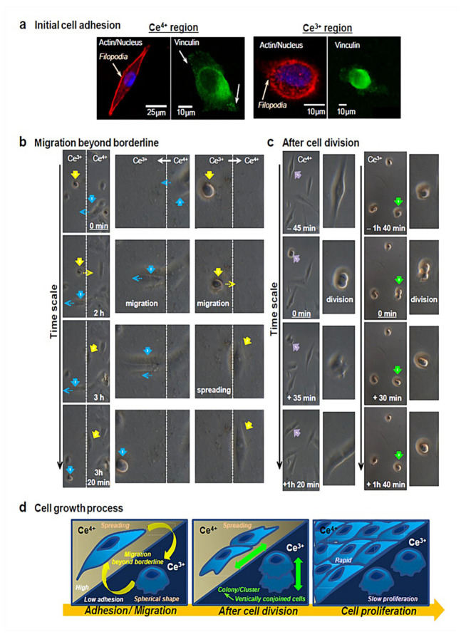
Figure 2.
(a) As for the assessment of initial cell adhesion in Ce4+ and Ce3+ regions, actin filaments (red), nucleus (blue), and vinculin localization (green) are observed by applying immunofluorescence staining to cells by confocal microscopy. Cells adhere to Ce4+ regions (A-IV) and Ce3+ regions (B-III). The development of actin filaments and vinculin localization is evident in Ce4+ regions, whereas Ce3+ regions are found with no actin filaments and vinculin localization in the spherical-shaped cells. Interestingly, filopodia are active in both regions to a great extent. Filopodia are responsible for sensing the cell environment and building new adhesion contacts that further trigger cell migration and spreading. Therefore, cells are able to attach to Ce3+ sites by activating filopodia on a plasma membrane. (b) Cells can migrate beyond the borderline at a timescale of around 3h. Accordingly, cellular migration comes about from Ce4+ to Ce3+ regions (blue arrows) and vice versa (yellow arrows), as evidenced by reversible changes in the cellular morphology between spread shapes and spherical shapes. (c) In order to evaluate cell proliferation, cell morphology prior to/following cell division is observed. Cell division takes place in Ce4+ regions (purple arrows), with cells attaching to the substrate surface as per normal and then spreading. By contrast, cells undergo division on Ce3+ regions (green arrows), present as one cell, implying that cells are vertically conjoined and scarcely migrate on Ce3+ regions. When at least 8 h pass, cell separation and attachment are seen in Ce3+ regions, followed by initiated cell migration. Therefore, cells scarcely attach to Ce3+ regions, which, in turn, eases the development of a cell cluster/colony and supports strong cell–cell interactions but weak cell–material interactions. (d) All the events, namely cell adhesion, cell morphology, and proliferation, involved in cell growth in Ce4+ and Ce3+ regions are summarized schematically. Reprinted with permission from [109].

