Figure 2.

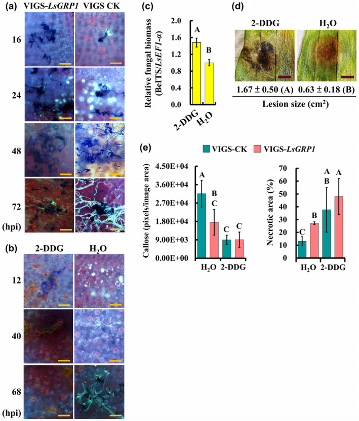
LsGRP1‐enhanced callose deposition suppresses Botrytis elliptica infection in lily. (a) LsGRP1 silencing reduced callose deposition in response to B. elliptica challenge. LsGRP1‐silenced (VIGS‐LsGRP1) and VIGS‐control (VIGS‐CK) leaves were droplet‐inoculated with a 20‐μl spore suspension of B. elliptica at 5 × 10⁴ spores/ml. Callose deposition and fungal growth were visualized by aniline blue and trypan blue double staining. hpi, hours post‐inoculation. (b)–(d) Inhibition of callose deposition in lily enhanced B. elliptica infection and symptom development. Lily leaves were droplet‐inoculated with a 20‐μl spore suspension of B. elliptica at 5 × 10⁴ spores/ml at 28 hr after infiltration with 1 mM 2‐deoxy‐d‐glucose (2‐DDG, a callose synthesis inhibitor) or sterile deionized water (H2O). (b) Effect of callose inhibitor on callose deposition and in planta fungal growth visualized by aniline blue and trypan blue double staining. (c) Relative B. elliptica biomass at 68 hpi detected by quantitative PCR. (d) Symptoms at 7 days post‐inoculation (dpi). (e) The treatment of 2‐DDG reduced LsGRP1‐conferred grey mould resistance. Lily leaves were spray‐inoculated with B. elliptica at 5 × 10⁴ spores/ml at 28 hr after 2‐DDG infiltration. Callose deposition and necrotic lesions were detected and quantified at 16 hpi and 3 dpi after aniline blue and trypan blue staining, respectively. Data represent the mean ± SD from three, three, and five biological replicates in (c), (d), and (e), respectively. Statistical analysis was performed using analysis of variance followed by LSD test (p < .05). Bar: 100 μm in (a) and (b); 0.5 cm in (d)
