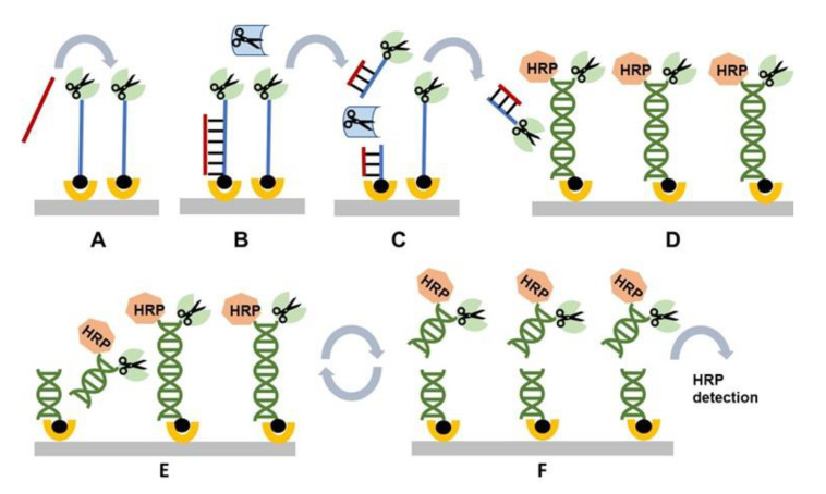
Figure 4.
General schematic of the restriction cascade exponential amplification (RCEA) assay. (A) An oligonucleotide probe specific for a target of interest is conjugated to an REase for amplification and attached to a solid substrate using biotin. A test sample containing the target of interest is added. (B) The target in the test sample hybridizes to the probe. (C) The hybrid is specifically cleaved by a recognition REase. amplification REase is subsequently released into the solution. (D) The reaction solution is transferred to an amplification cell that contains an excess of immobilized amplification REase attached to the surface through an oligonucleotide linker. The linker contains the restriction site corresponding to the amplification REase, and it is double-stranded, with the second strand conjugated with HRP. All amplification REase molecules in the amplification cell are immobilized and thus incapable of cleaving their own or neighboring linkers. Addition of the free amplification REase generated in (C) triggers linker cleavage, releasing additional amplification REase, which in turn cleaves new linkers. (E) Each step of this exponential cascade of cleavage reactions doubles the amount of free amplification REase molecules in the reaction solution. (F) The linker cleavage releases HRP, which is quantified colorimetrically. Each initial target–probe hybridization event produces an exponentially amplified number of HRP molecules, with the value dependent on the amplification time.

