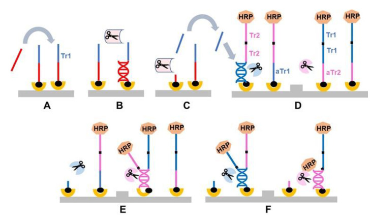
Figure 6.
General schematic of tandem oligonucleotide repeat cascade amplification (TORCA). (A–C) The recognition stage: An oligonucleotide probe specific for a target of interest is extended with a “trigger” unit (Tr1) and attached to surface using biotin. A test sample containing the target of interest is added (A). The target in the test sample hybridizes to the probe (B), and the hybrid is specifically cleaved by a specific recognition REase (C). The Tr1 unit is subsequently released into the reaction solution. (D–F) The amplification stage: The reaction cell carries two types of amplification probes. The first contains a single unit complementary to the trigger sequence Tr1 (antisense Tr1, aTr1), and multiple identical units of a trigger sequence Tr2. The second contains multiple identical Tr1 units, and a single unit complementary to the Tr2 unit (antisense Tr2, aTr2). Both probe types are surface-attached and contain a molecular marker HRP on their solution-facing end (D). The reaction solution in the amplification chamber contains two common REases, specific to Tr1 and Tr2, that recognize and cleave dsDNA hybrids of Tr1-aTr1 and Tr2-aTr2, respectively. When the recognition reaction solution is transferred to the amplification cell, the free trigger Tr1 hybridizes to an aTr1 unit of the first probe leading to the probe cleavage by Tr1-REase (D) and release of Tr2 into the reaction solution (E). In turn, the released Tr2 hybridize to an aTr2 of the second probe type (E), causing cleavage of Tr2 and further release of additional Tr1 units. This cascade of events also results in the release of the HRP molecular marker that can be used for signal quantification (F).

