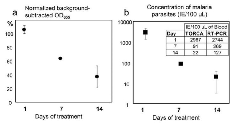
Figure 8.
From [10] (Creative Commons CC-BY license). The dependence of the TORCA signal (a) and the calculated infected erythrocyte concentration (b) on the time after initiation of the patient drug treatment (X-axis, days). (a) The Y-axis shows the background-subtracted and normalized HRP signal values. The background was calculated as the mean signal generated for the triplicate no-trigger added negative controls. For normalization and comparison of the sample series, the HRP signal values are expressed as the percentages of the background-subtracted signal obtained for the positive control containing an equimolar mixture of targets at 10 nM concentrations. (b) The Y-axis shows the IE (Infected Erythrocytes) concentrations calculated using the standard calibration curve obtained separately. The inset shows mean data for IE concentrations measured using TORCA and reverse transcription-PCR methods and calculated according to corresponding calibration curves. Error bars in both graphs show standard deviations. Error bars for 7-day treatment are smaller than the marker.

