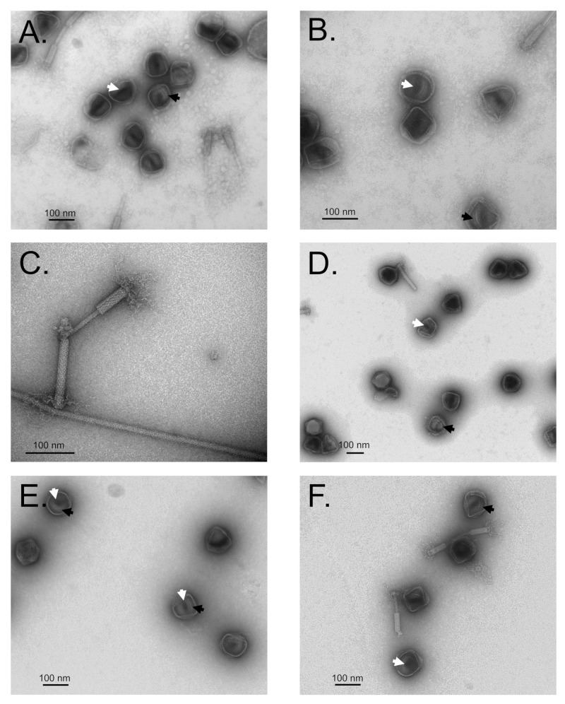
Figure 3.
Transmission electron microscopy of Salmonella phage SPN3US protease mutants 245(am59) (A–D) and 245(am66) (E,F). Many precursor head particles and tails were observed. Numbers of precursor head particles were observed to have internal material (black arrow) that contained a central darkly staining region (white arrows). This darkly staining region was interpreted to not be DNA as there was no detectable DNA in these samples (see Figure S2). (A,B,E,F) Particles treated with DTSSP prior to purification (no extensive crosslinking of major head proteins was observed by SDS-PAGE). (C,D) Particles were treated with glutaraldehyde prior to purification. (C) A contractile tail apparently cross-linked to a flagellum via its baseplate/fibers.

