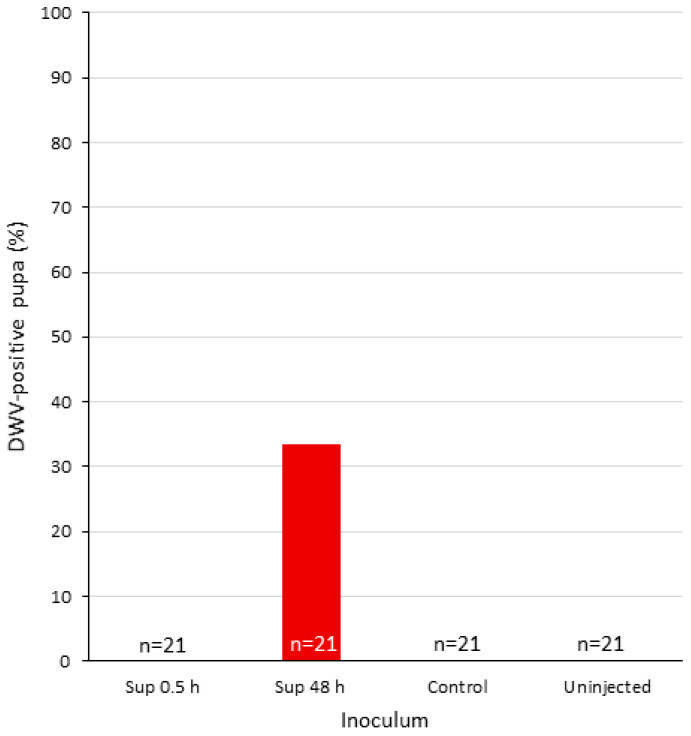
Figure 5.
DWV prevalence of injected pupae. X-axis, individual pupae treated as follows: Injected with DWV-P1 at 0.5 and 48 h (Sup. 0.5 and 48 h, respectively) supernatant from mock-infected cells (control) or uninjected (see Materials and Methods, Section 2.5).

