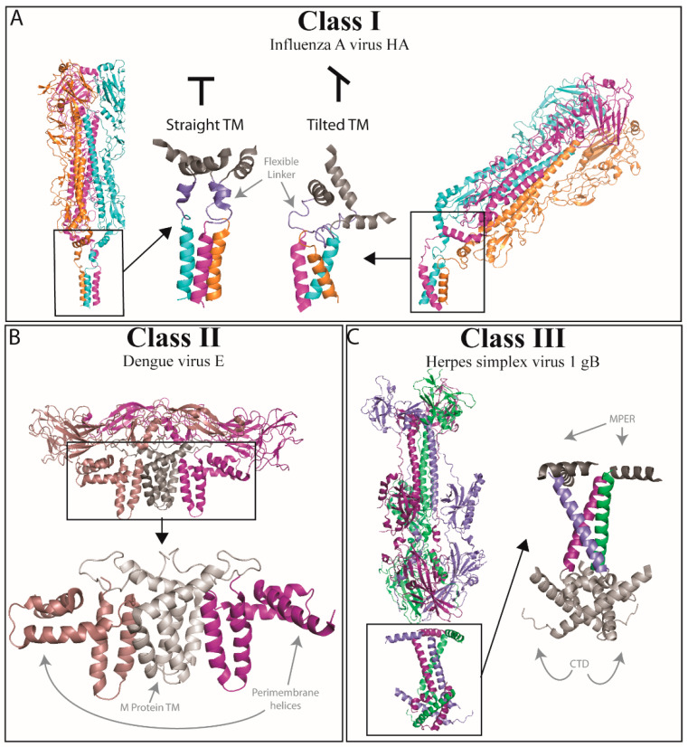
Figure 2.
Full-length structures of viral fusion proteins. While the ectodomain structures of numerous viral fusion proteins have been solved, only a few solved structures of full-length viral fusion proteins, including the transmembrane domain (TMD) have been solved. (A) Full-length influenza hemagglutinin (HA) was solved in 2018 [51]. Two structures were published, 6HJQ (far left) and 6HQR (far right). The first is HA in its pre-fusion conformation, and the ectodomain is in line with the TMD helices (middle left, zoomed in). In the second structure, the ectodomain is tilted 52° with respect to the TMD helices (middle right, zoomed in); this likely represents a scenario that is an intermediate state of fusion. In each zoomed-in area, a linker region is indicated (slate), and this region remains flexible to help compensate for this tilt. (B) In 2013, the structure of full-length Dengue virus E protein was solved [53]. This structure showed the hetero-tetramer complex of two E proteins (light and dark purple) and 2 M proteins (gray) with their respective TMD helices (3J2P). Flavivirus E proteins have two TMDs that have extensive hydrophobic interactions between them. The structure also includes three peri-membrane helices that lie on the outer surface of the viral membrane, approximately perpendicular to the TMD helices. (C) Full-length Herpes simplex virus 1 gB protein was published in 2018 [54]. This structure (5V2S) shows the three TMD helices situated in a triangular teepee structure; the MPER (dark grey) is a helix that lies almost perpendicular to the orientation of the TMD helices. The solved structure also includes a large portion of the cytoplasmic tail (CTD, light gray). Each CTD has two helices, the first of which is a small helix that links to a larger helix which then angles back towards the inner leaflet of the viral membrane. Because of the orientation, these CTDs may act as a clamp that assists in holding the gB TMDs in specific conformation, and the angle of the CTD may work in concert with the TMD helices to dictate the overall conformation of the protein. Figure made with PyMOL (Schrödinger®, Neo York, NY, USA).

