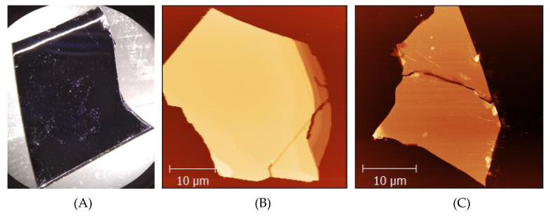
Figure 1.
An exemplary sample containing agglomerates of mostly thick mechanically exfoliated 2H MoS2 crystals on a silicon substrate. (A) An optical microscopy image of a typical sample being ca. 2 cm long and ca. 1.5 cm wide. Visible whitish spots are agglomerates of single MoS2 flakes. Two of such single MoS2 flakes are presented in (B,C). Image (B) is an AFM topograph of a flake with a mean height of a central zone (excluding several lone MoS2 pieces) of ca. 65 nm and a Z-scale of the image of 130 nm. (C) An AFM topograph of another flake with a mean height of a central zone of ca. 25 nm and a Z-scale of the image of ca. 65 nm. For images (B,C) brighter colors represent portions of the image, which are higher (Z-scale).

