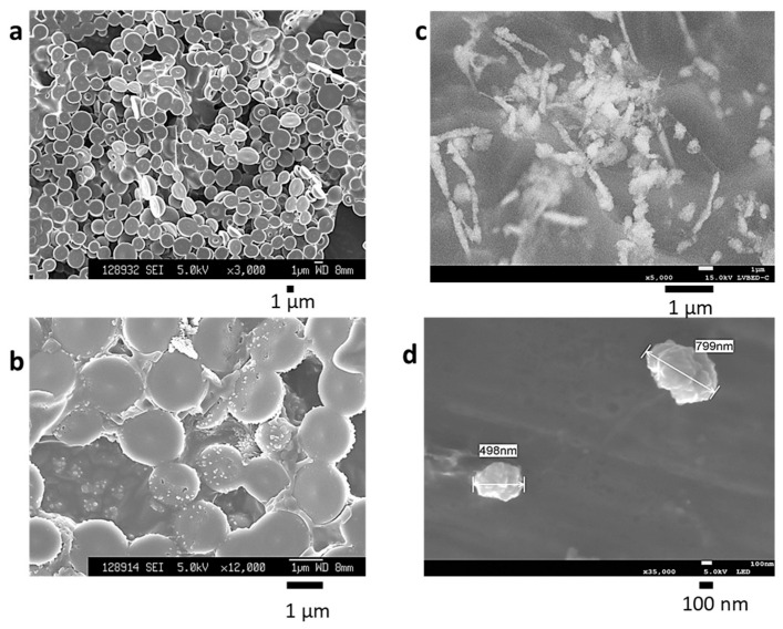
Figure 1.
Scanning electron microscopy (SEM) images of drying BiSCaO Water and cryo-SEM images of BiSCaO Water. The particle surface structure of drying BiSCaO Water at 3000-fold magnification (a) and at 12,000-fold magnification (b) was observed with SEM images taken with a field-resolved scanning electron microscope. Cryo-SEM observations were performed on BiSCaO Water at 5000-fold magnification (c) and 35,000-fold magnification (d). Arrows indicate nanoparticles comprising assemblies of smaller nanoparticles.

