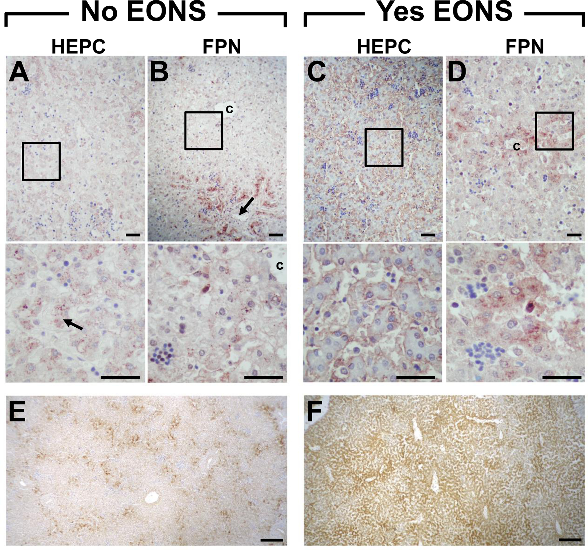
Fig. 4.

Representative immunohistochemical staining of liver hepcidin (HEPC, A&C) and ferroportin (FPN, B&D) of term newborns who died ~1 h postnatal. Liver of newborn who died of a non-septic cause (congenital diaphragmatic hernia, No EONS) shows (A) HEPC localized in hepatocytes in a granular and dispersed intracellular pattern (arrow), and (B) FPN staining more prominent around portal veins (arrow) and central vein. Liver of newborn who died of culture confirmed early-onset sepsis (Yes EONS) shows (C) intense HEPC staining lining sinusoids, and (D) diffuse FPN staining more conspicuous near central vein. Magnified insets of boxed regions are shown below the respective panel (A-D). Histochemical staining for liver iron stores for No EONS (E) and Yes EONS (F) sections shown in A-D. Original magnification: upper panels 200×; middle panels 600×; bottom panels 40×. Scale bar = 50 μm (A-D); scale bar = 200 μm (E&F); c = central vein.
