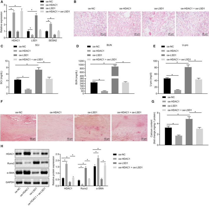
Figure 6.

Regulation of the HDAC1/LSD1/SESN2 pathway on VC in adenine‐induced CRF rats. A, The expression of HDAC1, LSD1 and SESN2 in thoracic aorta of rats determined using RT‐qPCR. B, HE staining of pathological changes of renal tissues (400×). C‐E, Serum levels of SCr, BUN, and U‐pro in rats measured by automated analyser Falcor 300. F, Alizarin red staining of calcification of thoracic aorta (200×). G, Calcium content in aortic tissue supernatant measured by colorimetric method. H, Western blot analysis of Runx2 and α‐SMA proteins in aortic tissues. BUN, blood urea nitrogen; CRF, chronic renal failure; GAPDH, glyceraldehyde‐3‐phosphate dehydrogenase; HDAC1, histone deacetylase 1; HE, haematoxylin‐eosin; NC, negative control; oe, overexpression; Pi, inorganic phosphate; RT‐qPCR, reverse transcription‐quantitative polymerase chain reaction; Runx2, Runt‐related transcription factor 2; SCr, serum creatinine; U‐pro, urine protein; VC, vascular calcification; VPA, valproic acid; VSMCs, vascular smooth muscle cells; α‐SMA, α‐smooth muscle actin. *P < .05 indicates significant difference. Data (mean ± SD) between two groups were analysed using unpaired t test. The experiment was run in triplicate
