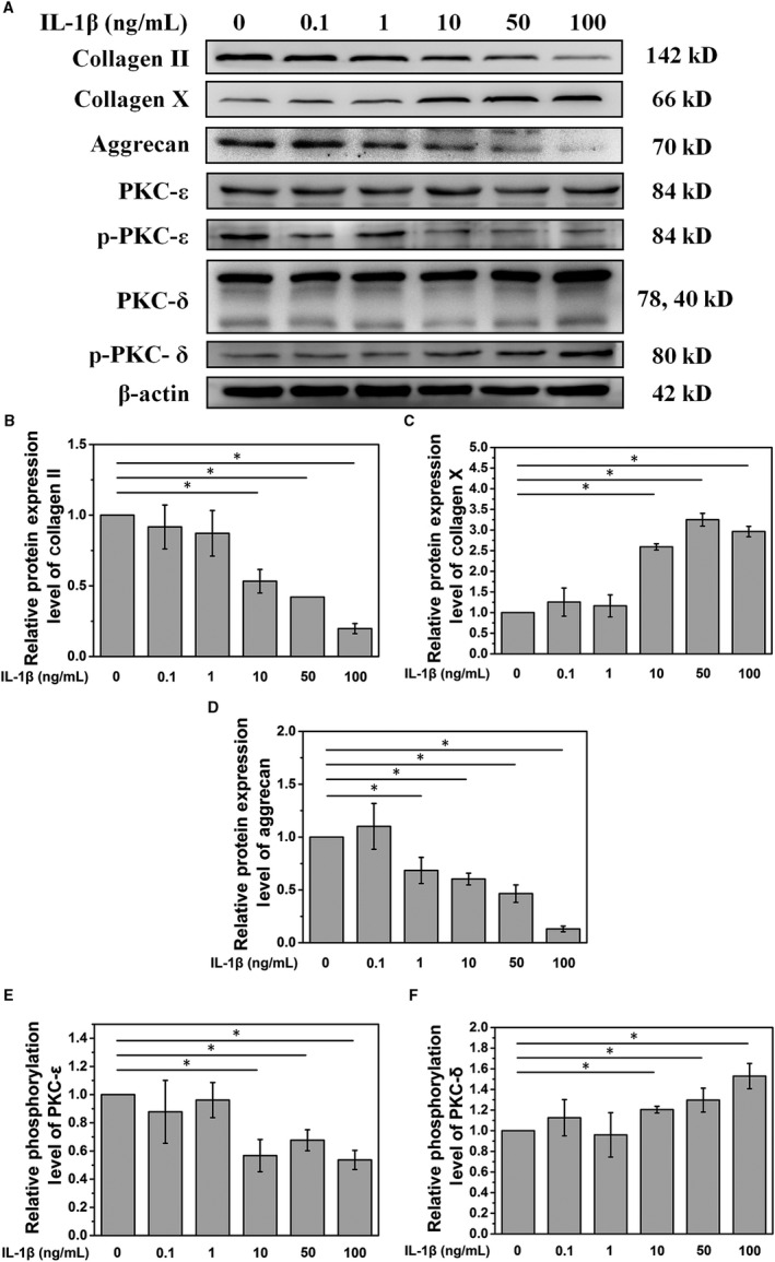
Figure 1.

A. IL‐1 treatment decreases PKC‐ε phosphorylation and increases PKC‐δ phosphorylation. B, C and D. Quantitative analysis of the Western blot showed that at a concentration of over 10 ng/mL, IL‐1 could significantly affect the expression of type II collagen, aggrecan and type X collagen. E and F. Quantitative analysis of the Western blot showed that a concentration of IL‐1 over 10 ng/mL could significantly affect PKC signalling (*P < .05)
