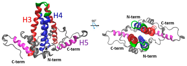
Figure 3.
Representation of the 3D structure of the HBc–NTD dimer. Two orthogonal views of the HBc dimer are shown. This dimer structure was obtained from the Protein Data Bank (PDB) 1QGT [24]. Each monomer is formed of five α-helices. Helices 3 (red) and 4 (blue) display a hairpin shape assembling into a four-helical bundle within the dimer, generating the capsid spikes of the particle. The spike tip (in green) corresponds to the highly immunogenic c/e1 epitope [23]. The fifth helix (magenta) and the downstream proline-rich loop are involved in the dimer–dimer oligomerization.

