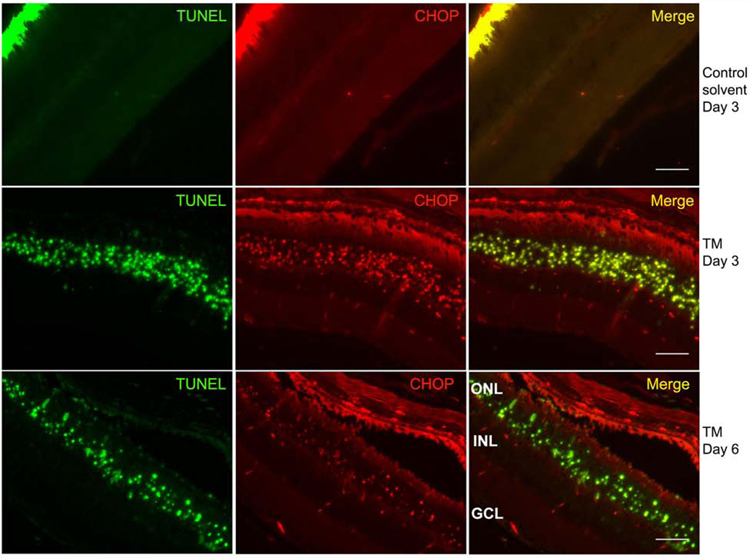
Fig. 6. CHOP expression in photoreceptor cells undergoing apoptosis.
Eye section from mice subjected to solvent control, tunicamycin treatments (0.1 µg) for 3 or 6 days were double stained with TUNEL (green) and CHOP (red). Tunicamycin treatment at day 3 (middle row) and day 6 (lower row) showed the apoptotic (TUNEL positive) and ER stress positive (CHOP positive) cells, which are located in the ONL. No staining was observed in sections from solvent control eyes (top row). Significant overlap staining is only seen in the ONL. (GCL: ganglion cell layer; INL: inner nuclear layer; ONL: outer nuclear layer). Scale bar, 100 µm.

