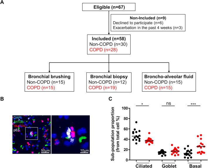
Fig. 1.
Airway progenitor basal cell population is enriched in COPD. a. Study flow chart. b. Representative micrograph showing a Region of Interest (ROI) containing AEC obtained by bronchial brushing in a non-COPD patient stained for cilia (Arl13b, green); mucins (Muc5ac, red); basal cells (p63, white) and cell nuclei (DAPI, blue). Magnification corresponding to the selected area is shown. C. Dot plot with median showing the percentage of ciliated, goblet and basal cells in both non-COPD (n = 15) (black circle) and COPD patients (n = 15) (red circle). *, p < 0.05 and ***, p < 0.0001; non-COPD vs COPD

