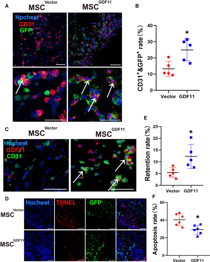
FIGURE 5.

Effect of GDF11 on MSC differentiation, retention and apoptosis in vivo. MSCGDF11 or MSCsVector expressing GFP or luciferase were mixed with Matrigel and implanted into mice. Paraffin sections of the recovered plugs 10 d after implantation were stained with DAPI for nuclei (blue) and specified antibodies (n = 5). A, Antibodies against GFP (green) and CD31 (red) were used for detection of endothelial‐like cells. White arrows point at the MSCs (orange) whose GFP was colocalized with CD31. The pictures in upper panel were taken at 400× magnification and the lower panel are magnified 2×. Scale bars: 50 μm. B, Rate of MSC differentiation into endothelial‐like cells was quantified by dividing number of double positive cells by number of total retained MSCs in an image. C, Antibodies against GDF11 (red) and CD31 (green) for endothelial‐like cells were used. White arrows point at the double positive cells (orange). The pictures were taken at 600× magnification. Scale bars: 50 μm. D, Sections were stained for TUNEL to identify apoptotic cells (red) and retained MSCs (GFP). Scale bars: 100 μm. E, Retention rates were quantified as the number of GFP+ cells out of the total number of cells. F, Apoptosis rate was quantified by the percentage of cells positive for TUNEL staining. Data are presented as the mean ± SD for at 2 independent experiments and were analysed. *P < 0.05; **P < 0.01 and ***P < 0.001
