FIGURE 3.

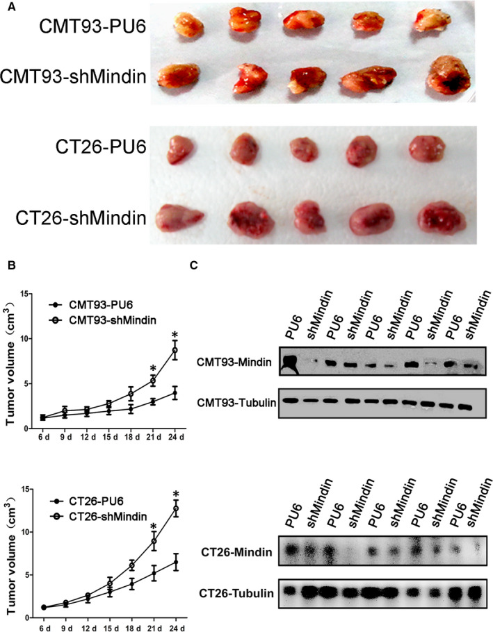
Subcutaneous implantation tumour growth of mindin knock‐down CMT93 and CT26 WT cells. C57BL/6 and BALB/c mice were subcutaneously injected with stable mindin knock‐down CMT93 or CT26 WT cells, or PU6 control cells. Tumour size was measured every 3 d for 24 d. A, Images of isolated tumours from the four groups of study mice (n = 5). B, In vivo tumour growth resulting from the mindin knock‐down CMT93 or CT26 WT cell (n = 5, *P < 0.05). C, Western blot analysis confirming mindin protein deficiency in the tumour tissues of the four study groups at the end of the study period. Tubulin was used as a protein loading control (n = 5). Upper panel indicates CMT93, and lower panel indicates CT26 WT among A‐C
