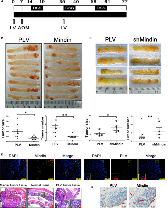
FIGURE 4.

Lentivirus‐mediated colitis‐associated cancer model. A, Experimental protocol used to induce CAC and the administration of lentiviral vectors. B, Images of isolated colon tissue from the mindin‐overexpression groups and control mice at the end of the study period (upper panel, n = 8), and tumour size and number (lower panel, n = 8, *P < 0.05, **P < 0.01). C, Photograph of isolated colon tissue from the mindin knock‐down groups and control mice (upper panel, n = 4), and tumour size and number (lower panel, n = 4, *P < 0.05, **P < 0.01). D and E, Confocal microscopy and anti‐mindin immunohistochemistry analysis of frozen and paraffin‐embedded colon sections showing the GFP reporter for lentiviral vector expression and mindin protein (as shown by the red arrows), and H&E staining of serial sections of mouse CAC tissues
