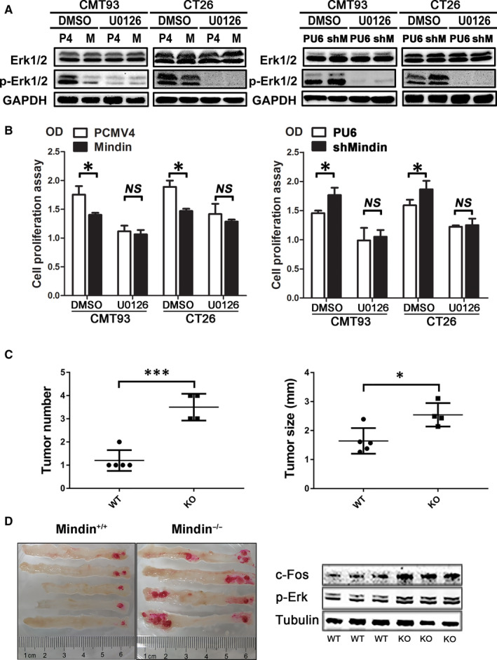
FIGURE 6.

U0126 inhibition of ERK1/2 phosphorylation, cell proliferation and colitis‐associated cancer model of mindin‐knockout mice. A, Western blot analysis of U0126‐treated cells using antibodies against ERK1/2 and phospho‐ERK1/2. GAPDH was used as a loading control. B, Analysis of U0126‐treated cell proliferation in the mindin‐overexpressing (left panel) or knock‐down (right panel) and control cells by BrdU assay (*P < 0.05). C, Tumour number (left panel) and size (right panel) of isolated colon tissue from the mindin‐knockout groups and control mice at the end of the study (n = 8, *P < 0.05, ***P < 0.01). D, Representative images of isolated colon tissue from the mindin‐knockout groups and control mice at the end of the study (left panel). Western blot analysis of the phosphorylation level of ERK in mindin‐knockout or control tumour tissues from CRC model mice (right panel). Tubulin was used as a loading control
