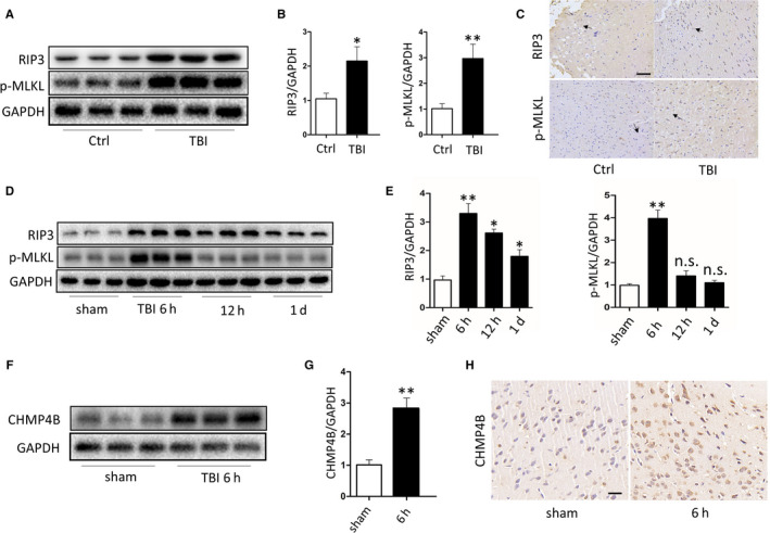
FIGURE 1.

There is necroptosis after traumatic brain injury and its level changes with time. A, Protein levels of RIP3 and p‐MLKL obtained from epileptic patients and severe trauma patients. GAPDH was used as the loading control. B, Bar graphs show the results of analysis (by band density analysis) of RIP3 and p‐MLKL (trauma patients = 3, epileptic patients = 3; data are presented as the means ± SEM). C, Immunohistochemistry of RIP3 and p‐MLKL in TBI and Ctrl. (Scale bar = 100 μm.) D, Protein levels of RIP3 and p‐MLKL obtained from sham mice and TBI mice at 6 h, 12 h and 1 d. GAPDH was used as the loading control. E, Bar graphs show the results of analysis (by band density analysis) of RIP3 and p‐MLKL (TBI = 5, sham = 5; data are presented as the means ± SEM). F, Protein levels of CHMP4B obtained from sham mice and TBI mice at 6 h. GAPDH was used as the loading control. G, Bar graphs show the results of analysis (by band density analysis) of CHMP4B (TBI = 5, sham = 5; data are presented as the means ± SEM). H, Immunohistochemistry of CHMP4B in sham and TBI at 6 h. (Scale bar = 50 μm.) *P < .05; **P < .01
