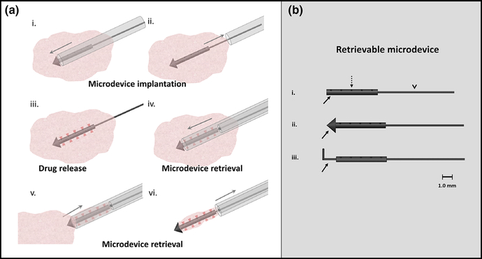
FIG. 1.
(a) Percutaneous microdevice implantation and retrieval schematic. An implantation needle delivers a microdevice into in-vivo tumor tissue (i) and (ii). The microdevice releases drug into ten spatially discrete sites over 24 h (red shaded regions, (iii). This tissue and the microdevice is percutaneously retrieved intact using an over-the-wire retrieval device to assess drug response (iv)—(vi). Up to ten drug combinations can be simultaneously assessed with a single microdevice. (b) (i) Retrievable microdevice with body (solid arrow) that contains ten distinct drug release reservoirs/sites (dashed arrow) preloaded with micro-doses of drug compound and released into adjacent tissue. A nitinol wire (arrowhead, v) attached to the back end of the microdevice body allows precise alignment and localization during retrieval. (ii) A “tophat” conical distal end (arrow) prevents microdevice migration after tissue implantation. (iii) A nitinol wire ‘anchor’ (arrow) can also be used to secure the microdevice in place.

