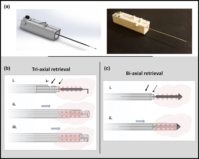
FIG. 2.
Microdevice retrieval prototype. (a) CAD model and actual prototype of a device allowing precise retrieval of the microdevice and a small adjacent cylindrical tissue sample. (b) Tri-axial retrieval device has an inner stylet (dotted arrow) that passes over the wire, a notched coring needle (solid arrow), and an outer end-cutting needle (arrow head). The retrieval device is advanced over the guidewire to the proximal end of the microdevice (row i), the coring and end cutting needles are advanced around the microdevice to cut and enclose the surrounding tissue (row ii) and the end cutting needle is advanced through the notch to completely sever the distal end of the tissue (row iii). (c) Bi-axial retrieval device has an inner stylet (dotted arrow) and outer coring needle (solid arrow). This device is advanced over the wire to the proximal end of the microdevice (row i) and the coring needle is advanced to cut/enclose and simultaneously sever the surrounding tissue (row ii).

