FIG. 3.
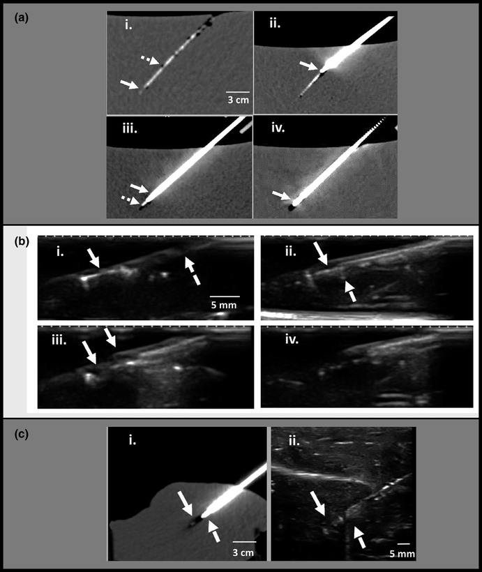
Computed tomography (CT) and ultrasound guided microdevice retrieval in phantom and ex-vivo liver tissue. (a) CT-guided retrieval in tissue-mimicking phantom. (i) Microdevice is implanted at a depth of 12 cm below the surface of a tissue-mimicking phantom. The microdevice (solid arrow) and nitinol wire (dashed arrow) are faintly seen. (ii) The retrieval tool is inserted into the tissue over the wire with a mild bend at the tip (solid arrow) indicating slight axis misalignment. (iii) Retrieval tool trajectory is corrected and advanced to the edge of the microdevice. The thicker coring needle (solid arrow) and thinner inner stylet (dashed arrow) are well seen. (iv) Coring needle advanced around the microdevice (arrow). (b) Ultrasound guided retrieval in tissue-mimicking phantom. (i) Microdevice is well visualized (solid arrow), and the wire is faintly seen (dashed arrow). (ii) retrieval tool is advanced and ultrasound confirms that its tip (dashed arrow) is sitting against the microdevice (solid arrow) with good alignment. (iii) the outer coring needle (dashed arrow) has been advanced halfway over the microdevice to cut the surrounding tissue. (iv) after tissue cutting, the entire retrieval tool with enclosed tissue is retracted and removed. (c) Ex-vivo animal tissue experiments demonstrate similar imaging findings as the phantom. (i) retrieval needle (dashed arrow) advanced to the edge of the microdevice (solid arrow) under CT guidance to depth of 10 cm. (ii) ultrasound image during tissue retrieval also shows the retrieval needle (dashed arrow) located at the edge of the microdevice (solid arrow) with good alignment.

