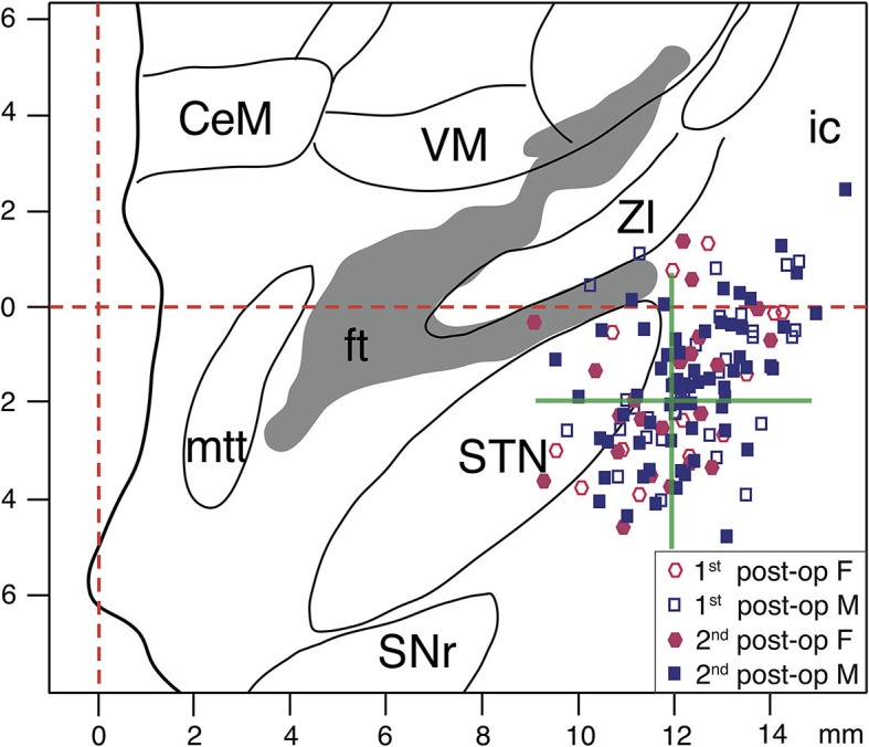
Figure 7.

Stereotactic reconstruction of active DBS contacts. The active DBS contacts for both hemispheres are superimposed on a frontal section of the stereotactic atlas of Morel et al., at mid-commissural point level (69). Empty and filled circles represent the active contacts for female patients at the 1st and 2nd post-operative stages, respectively. Empty and filled squares represent the active contacts for male patients at the 1st and 2nd post-operative stages, respectively. The interrupted red lines indicate midline and AC-PC level. The green cross represents the averaged stereotactic target coordinates aimed for the study group. In patients with two adjacent active contacts (cathodes), the averaged x, y, z stereotactic coordinates of those pairs have been plotted. CeM, centralmedialnucleus; ft, fasciculus thalamicus; ic, internal capsule; mmt, mammillothalamic tract; SNr, substantia nigra, pars reticulata; STN, subthalamic nucleus; VM, ventral medial nucleus; ZI, zona incerta.
