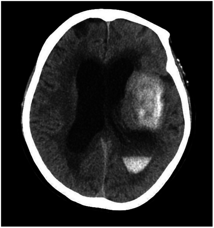
Figure 10.
Twelve hours after the scan in Figures 4–7, repeat head computed tomography indicated acute hydrocephalus with significant enlargement of the fourth ventricle, third ventricle, and bilateral lateral ventricles. The high-density shadow in the basal ganglia, occipital horn of the left lateral ventricle, and frontal lobe had also become lighter.

