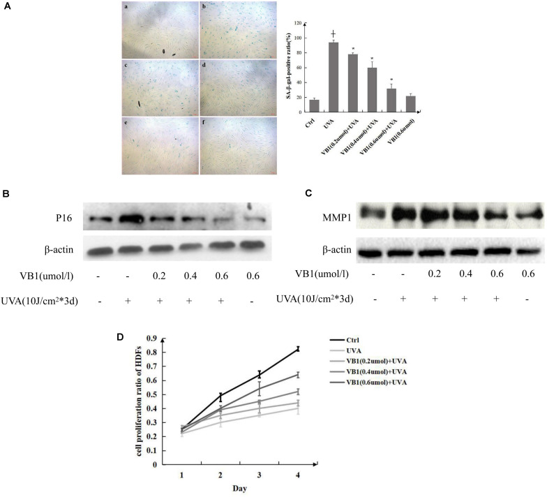
FIGURE 1.
VB1 protects HDFs from UVA-induced senescence. (A) HDFs senescence was evaluated by measuring the number of SA-β-gal-positive cells (left panel) [a, Ctrl; b, UVA; c, VB1 (0.2 μmol/l) + UVA; d, VB1 (0.4 μmol/l) + UVA; e, VB1 (0.6 μmol/l) + UVA; f, VB1 (0.6 μmol/l)]. Percentages of SA-β-gal-positive cells were determined by counting stained cells and total cells in four continuous visual fields under a microscope (200x). The SA-β-gal-positive rate was obviously enhanced in UVA-induced HDFs, while VB1 inhibited UVA-induced SA-β-gal activity in a dose-dependent manner. The analysis data was shown in right panel. Data are presented as mean HDFs ± SD (n = 3; + vs. ctrl, p < 0.05, * vs UVA, p < 0.05). (B) p16 levels were detected by western blot analysis. Cells were irradiated as described and harvested 24 h after the final UVA exposure. Blots were probed to detect p16, stripped, and then reprobed for β-actin. VB1 inhibited UVA-induced p16 expression in a dose-dependent manner. Images are representative of 3 independent experiments. (C) MMP1 levels were detected by western blot analysis. VB1 inhibited UVA-induced MMP1 expression in a dose-dependent manner. Images are representative of 3 independent experiments. (D) The HDFs growth rate was determined by performing MTT assays. The growth rate of UVA-exposed HDFs was significantly decreased compared with control HSFs, while VB1 could reverse the decrease (n = 3 for each time point).

