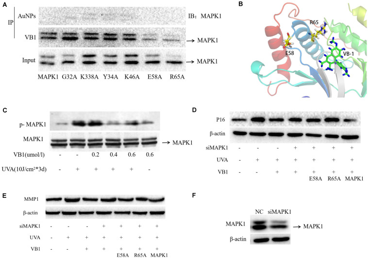
FIGURE 4.
VB1 binds to MAPK1 through E58 and R65 residues of MAPK1 and VB1 protects 293T cells from UVA-induced senescence via binding the two residues. (A) VB1 linked with gold nanoparticles was used for the immunoprecipitation in 293T cells. In the gold nanoparticles (AuNPs) immunocomplexes, MAPK1 was not detectable by western blot analysis, while MAPK1 was detectable by western blot analysis in nanogold-VB1-immunoprecipitated complexes. In the 293T cells transfected with E58-mutant MAPK1 and R65-mutant MAPK1, MAPK1 was not detected, while MAPK1 was detectable in cells transfected with other four mutant MAPK1. (B) The combination form between VB1 and MAPK1 via E58 and R65 residues using computer-aided methods. (C) p-MAPK1 expression was detected by western blot analysis. p-MAPK1 was significantly increased after UVA irradiation and that VB1 could significantly decrease UVA-induced p-MAPK1 expression in a dose-dependent manner. (D,E) 293T cells was used in further co-transfected experiments because of difficulty in HDFs. Endogenous MAPK1 in 293T cells was knockdown by MAPK1 siRNA. Then UVA-irradiated 293T cells were co-transfected with VB1 and wild-type or mutant MAPK1 (E58-mutant or R65-mutant), and p16 and MMP1 expression was detected by western blot analysis. UVA-induced p16 and MMP1 were significantly decreased in 293T cells tranfected with wild-type MAPK1 and VB1, while p16 and MMP1 were partially reversed in 293T cells transfected with E58-mutant or R65-mutant MAPK1 and VB1. (F) MAPK1 siRNA could significantly knockdown the endogenous MAPK1 expression in 293T cells.

