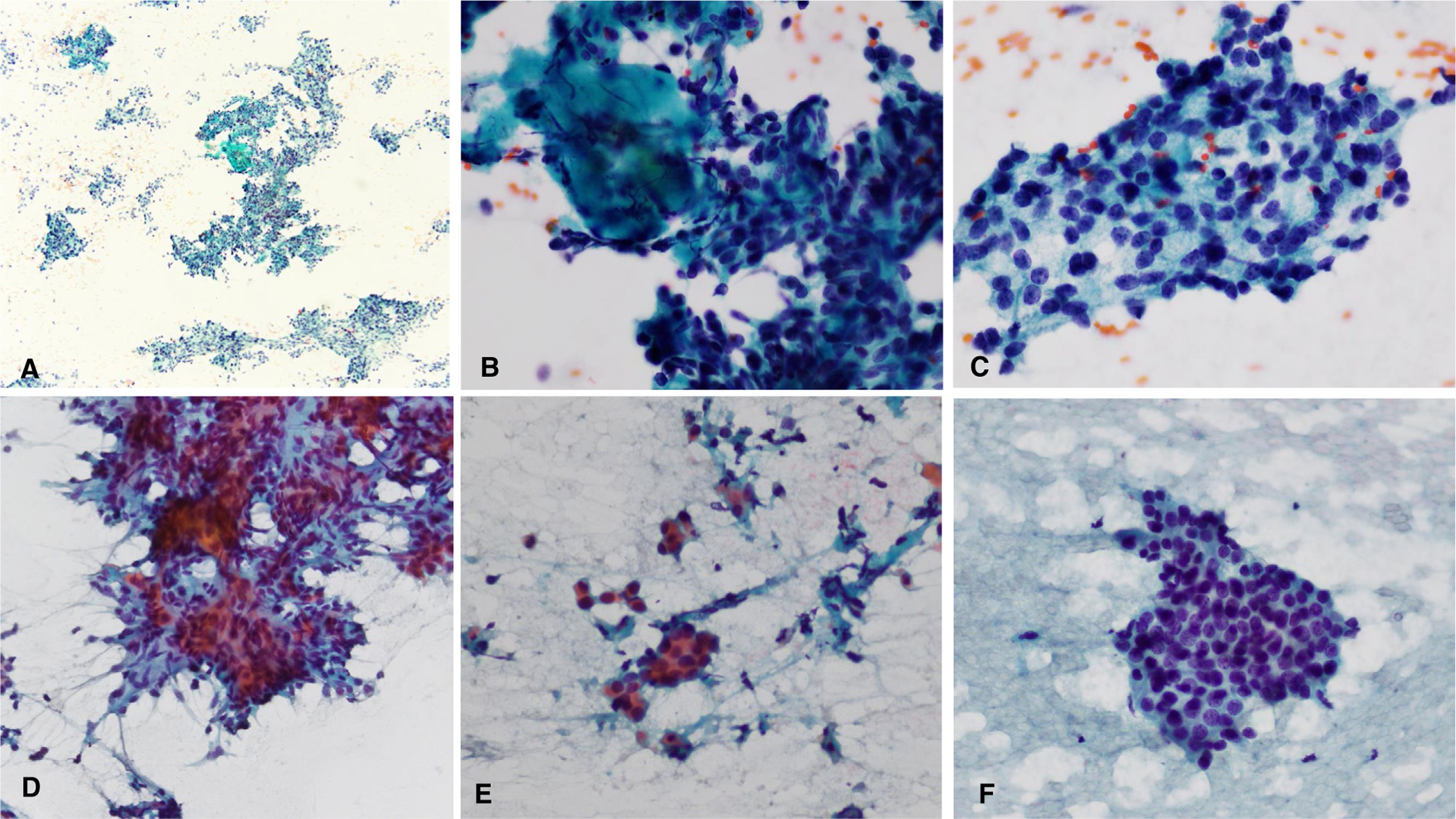
FIGURE 3.

Two difficult low-grade salivary gland carcinoma cases. (A-C) Acinic cell carcinoma with hypercellularity with crowded groups, focal fibrous tissue, and variable hyperchromasia that likely led to overgrading. (D-F) Epithelial-myoepithelial carcinoma with hypercellularity, small crowded groups, background debris, and mild hyperchromasia that likely were problematic for accurate grading. (Papanicolaou stain, A, original magnification ×40, B-F, original magnification ×400.)
