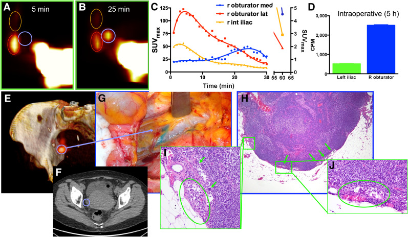
FIGURE 1.
Representative example of clinical PLG with PET/CT and intracervical injection of 18F-FDG in patient with uterine carcinosarcoma (patient 1). (A–C) PLG demonstrated 3 SLNs in right pelvis (A and B), 1 of which demonstrated a prolonged and delayed uptake pattern, as shown in time–activity curves (C). (D) Intraoperative measurements of counts per minute (CPM) of excised LNs demonstrated, even 5 h after tracer injection, higher uptake in suggestive node. (E and F) Three-dimensional surface reconstruction of PET/CT at 25 min showing higher uptake in suggestive node (blue circle, E) and same node as shown on axial CT (F). (G) Intraoperative view of node stained by vital blue dye. (H–J) Low-magnification (H) and high-magnification (I and J) histologic photomicrographs confirming presence of metastatic carcinoma within suggestive node.

