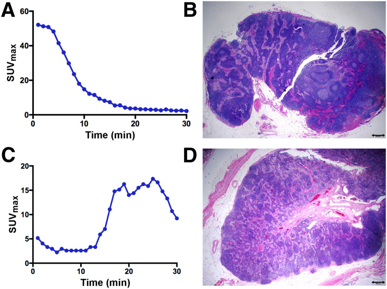
FIGURE 3.
True-negative and false-positive PLG. (A and B) Patient 23 with high-grade endometroid adenocarcinoma and left obturator SLN that was negative on PLG (A) and pathology (B). (C and D) Patient 11, with high-grade endometrial cancer, showing unilateral PLG mapping with prolonged increase and delayed peak of 18F-FDG uptake (C) within right external iliac SLN, which was suggestive of malignancy; this finding corresponded to right external iliac SLN at time of surgery (concordant mapping), but final pathology revealed this SLN to be negative for tumor cells (D).

