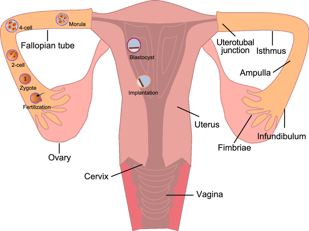
Figure 1.
The anatomy of human female reproductive system and early pregnant events. The female reproductive system consists of the ovaries, fallopian tubes, uterus, cervix, and vagina. Following fertilization in the fallopian tube, the zygote undergoes 5–6 miotic cell division to form morula, a 16-cell embryo. Simultaneously, the developing embryo continues to be projected toward the uterus in the fallopian tube and reaches the uterus approximately 3 days after fertilization. Once inside the uterus, the morula continues to divide and produce an approximately 100-cell embryo, which called blastocyst. The blastocyst consists of two major structures: the mass of cells inside of blastocyst is called inner cell mass (ICM) which will become the embryo; the cells that form the outer shell of the blastocyst are trophoblasts which will develop into chorionic sac and fetal portion of placenta. The trophoblasts of blastocyst then secrete enzymes to degrade zona pellucida, which is referred to as hatching. At about day 7 after fertilization, the hatched blastocyst adheres to the uterine endometrium for embryo implantation, which is complete at about day 10 after fertilization.

