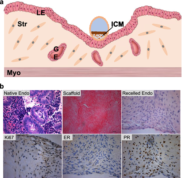
Figure 4.
(A) Uterine anatomy and embryo implantation. The blastocyst initiates the implantation process through embryo apposition, adhesion, and penetration to the uterine luminal epithelium (LE). The LE cells around the attachment site will start apoptosis upon blastocyst attachment, and help the blastocyst penetrate the LE layer into the stroma. ICM: inner cell mass. Str: stroma cells. GE: glandular epithelium. Myo: myometrium. Modified based reference [91]. (B) Bioengineering model of human uterine endometrium. Human endometrium tissue before and after the decellularization, after recellularization, and immunohistochemistry staining of Ki67, estrogen receptor (ER), and progesterone receptor (PR) on day 14 (28 days in microfluidic culture) in EVATAR.

