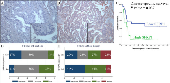
Figure 5.
Immunohistochemistry staining of SFRP1 in ampullary adenocarcinoma. (A) Negative expression of SFRP1. (B) High expression of SFRP1. (C) Kaplan–Meier analysis of the impact of SFRP1 expression on disease-specific survival in ampullary adenocarcinoma patients (P = 0.037). (D) Correlation between localisation of E-cadherin staining and expression of SFRP1 in using immunohistochemistry (IHC). High expression of SFRP1 is slightly correlated with the loss of E-cadherin, and low expression of SFRP1 is correlated with membranous staining of E-cadherin (P = 0.362). (E) Correlation between the localisation of β-catenin staining and expression of SFRP1 using IHC. High expression of SFRP1 is slightly associated with membranous staining of β-catenin, and low expression of SFRP1 is associated with nuclear staining of β-catenin (P = 0.301). IHC, immunohistochemistry; SFRP1, secreted frizzled related protein 1.

